Internal invivo radiation measurement to give clinicians confidence that doses are delivered to plan
The Technology
DOSEmapper™ is a unique high performance thermoluminescent detector (TLD) array consisting of 100+ small (0.95 – 1.6 mm) micro silica beads arranged on a thread for use in a standard catheter and inserted into a natural orifice next to the tumour. The inserted beads absorb the received radiation as stored energy over 100 points across and around the tumour. This is applicable to 70% of cancer occurrences.
After the radiotherapy session (usually the first fraction), the dose levels and spread will be measured by our fully automated high-performance TLD reader. The results will be ready, typically, after 20 minutes and sent to a local computer connected by USB. The measurements are a unique “radiation ruler” or profile of the actual radiation doses received along or across the tumour area and beyond at a resolution of ±1 mm.
The clinical team can see the beads, their sequence and positioning on the patient imaging system and can compare the measurements provided with those expected with the patient treatment plan. The team can decide if adjustments need to be made for the next session.
Current Problems with Radiotherapy
DOSEmapper™ enables clinicians to see and measure where radiation actually hits at and around the target tumour
Radiotherapy in Practice
Radiotherapy clinical teams carefully calculate and pre-test radiation treatment for each cancer patient. For external beam therapy the calculated radiation beams are directed at the tumour from the best and safest angles. Great care is taken to minimise hitting the surrounding healthy tissue and especially the patient’s critical organs. But, despite all the care, between 10% and 25% of radiotherapy patients worldwide suffer serious side effects or treatment failure.
One problem is how to directly measure the actual radiation dose delivered into the patient’s body during radiotherapy. And that includes in brachytherapy when radiation sources are inserted into the patient. Today no one can see what the radiation hits or if it hits solely according to the specified patient treatment plan. No one knows the effects until the patient reports problems some weeks later.
TRUEinvivo® micro silica bead array detectors are easily inserted via a catheter into a suitable natural orifice and measure the dose and spread of the radiation received at the site of the tumour. The clinical team can use this “radiation ruler” to check for accuracy against the plan immediately after the first patient session and, if necessary, adjust for the next dose. This test and correction for accuracy gives the clinical team real confidence that the correct dose has been given and in the correct places. More confidence can lead to less sessions.
The patient and clinicians still wait a few weeks to see if the cancer is cured but the wait is with the confident knowledge that the treatment was really accurate and that side effects and harm have been minimised.

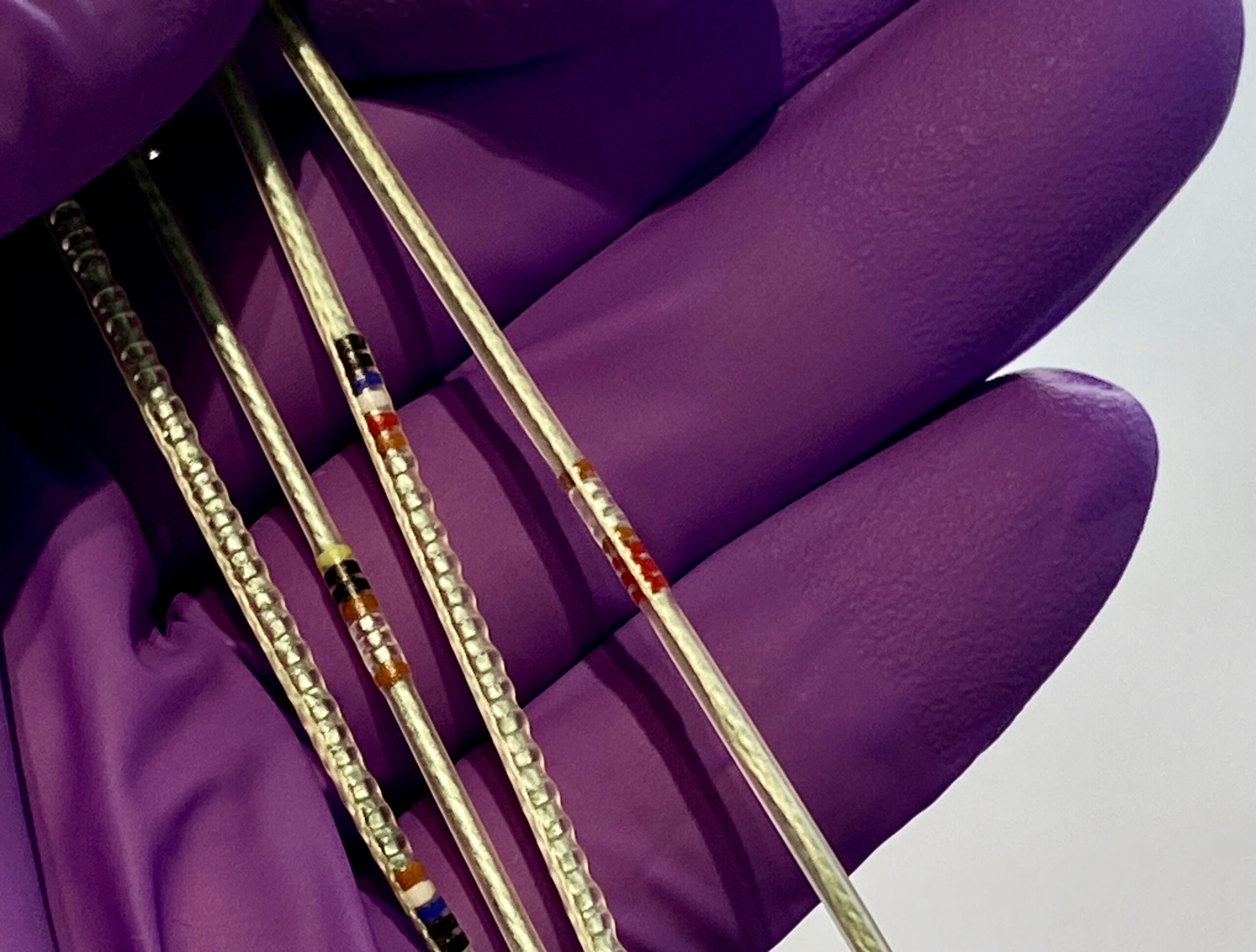
DOSEmapper1Ds™ on a gloved hand: for direct internal invivo dose measurement


DOSEmapper™ (micro silica bead TLD arrays)
TRUEinvivo 1D (linear), 2D(mesh) and 3D (cavity) DOSEmappers™ are suitable for medical physics applications that use X, γ, β, proton and neutron radiations. This includes external beam radiotherapy and brachytherapy treatments. For more details of the actual TLD material see the Researcher pages.
The DOSEmapper1Ds™ shown on the glove above are examples of bead arrays used for a very high resolution dose profile measurements in any natural orifice. The DOSEmappers™ are typically used for the planning CT and the first fraction. Our automated reader reads the DOSEmapper™ 160 TLDs in about 30 minutes. DOSEmapper1Ds™ can be ordered to any length and different spacing/resolution. We have supplied them with as few as 3 detector beads at 1mm resolution for commissioning work.
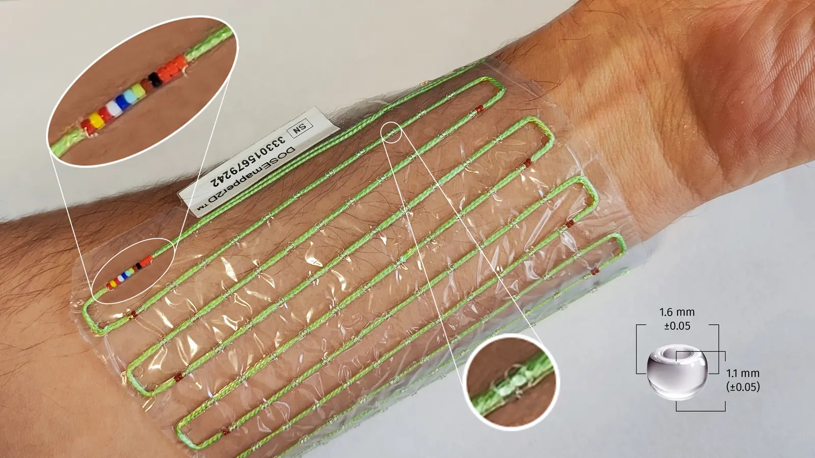
The DOSEmapper2D™ shown on the left (10 x 10 taped to the arm) has 100 double bead measuring points in a mesh that can be used to obtain surface dose measurements over any flat or curved skin area. The DOSEmapper2Ds™ also come in 5 x 5 and 20 x 20 sizes. Other sizes can be made to order.
The DOSEmapper3D™ (prototype also shown on the left) effectively has a DOSEmapper1D™ wrapped in a spiral along the length of a central catheter and former. When used as a rectal insert the DOSEmapper3D™ measure the received doses at the rectal wall. This is especially useful for the treatment of prostate cancer when adaptive treatment or hypofractionation is intended. In hypofractionation, the total number of fractions could be reduced if the doses to organs at risk could be measured giving clinicians confidence to safely increase the fractional doses.


